 English
 中文
中文 Français
 Español
 Pусский
Application of biopsy needle guide in clinical ultrasound intervention
1. Summary: Advantages of Ultrasound Intervention
Interventional medicine is a new diagnostic and treatment technology developed in recent years with the continuous advancement of imaging technology. It is a new medical branch besides internal medicine and surgery. The three technologies are combined to treat diseases and save patients. Its emergence enables a faster development of modern medical technology. Some previously incurable diseases have become curable, and some previously painful diseases have been treated with little or no pain. Due to so many advantages of interventional technology, it’s been advancing rapidly. Interventional ultrasound is an important part of interventional technology. Its safety, accuracy, effectiveness, and wide applications have put it in a leading position in modern medicine. It is easy to operate, low cost, less painful, and reliable in efficacy and becomes increasingly popular.
Injections and transfusions are something that people take for granted. Interventional ultrasound is actually a technique of "needling" under ultrasound guidance. Therefore, its operation is not only accurate, safe and secure, but also has minimal trauma and leaves no traces on the skin after surgery. Practice has proved that interventional ultrasound has solved problems that seems impossible by internal medicine and surgery, and it’s replaced some of surgical methods or as good as surgical operations. 【1】
2. Clinical application of ultrasound intervention
The application range of interventional ultrasound, with the improvement of ultrasound technology and the deepening of the understanding of ultrasound in various clinical departments, has been continuously expanded in both longitudinal and lateral aspects. In terms of scope, it includes the following commonly used applications.
• Fluid Aspiration: pleural fluid, amniotic fluid, and eggs retrieval;
• Needle biopsy: sampling of occupying lesions of liver and pancreas, etc.;
• Biospy imaging: gallbladder, renal pelvis, seminal vesicles, etc.;
• Biopsy: biopsy of liver, pancreas, kidney, prostate, intra-abdominal masses, intra-pelvic masses and retroperitoneal masses;
• Medicine injection and blood transfusion: sclerotherapy of liver and kidney cysts, hydatid cyst sclerotherapy and ovarian cyst sclerotherapy, liver Ca injection of alcohol, seminal vesicle injection of antibiotics for chronic seminal vesicles, fetal blood transfusion;
• Introduce physical factor therapy: liver cancer nuclide yttrium-90 glass microsphere injection, liver cancer microwave therapy, liver cancer cryotherapy, prostate afterloading therapy, high-intensity focused ultrasound therapy;
• Ultrasound surgery: cataract lens emulsification, liver cancer resection, kidney cancer resection, ultrasound liposuction (slimming);
•Intracavitary ultrasound: rectal ultrasound, vaginal ultrasound, esophageal ultrasound, narrow-diameter catheter ultrasound, used for intravascular, endoscopy, urethra, and uterine cavity;
• Intraoperative ultrasound: intraoperative ultrasound or needle biopsy of abdominal organs, intraoperative neurosurgery examination.
3. Interventional application of biopsy needle guide in various clinical directions
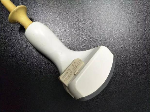
In the application of interventional ultrasound, besides various biopsy tools, the most important are ultrasound imaging machine and ultrasound probes with puncture guides or central groove-type puncture probes. Traditional puncture probes are mostly linear array probes, with a needle notch in the center of the probe, and there is no crystal (or element) at the needle groove notch. In the ulrasound image, a strip-shaped dark band is formed as a guide line, and the dark band is aligned during operation to the target, the puncture needle penetrates the target along the dark belt through the gap of the needle groove. Since the puncture needle is inserted in the dark belt, it is impossible to observe the location of the needle in real time, and it is basically not used now (Pic 1).
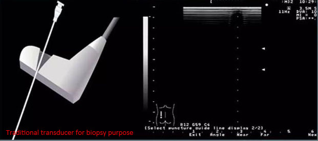
Pic 1 Conventional transducer for biopsy purpose
However, with the development of ultrasound technology, many ultrasound manufacturers have developed special abdominal puncture probes. Although some crystal (or element) of acoustic head are missing, the image will remains complete, thus making up for the shortcomings of traditional puncture probes. The following convex array puncture probes are from GE, Hitachi, Toshiba, ESAOTE (see Pic 2):
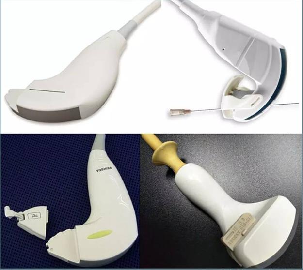
Pic 2 Abdominal-puncture transducers
At present, the most common ultrasonic intervention application is to mount a biopsy needle guide to the ordinary conventional probe (Pic 3), but some special probes with the biopsy needle guide can also be extended to more in-depth puncture applications (4D abdominal probe RAB6-D, Pic 4) 4D intracavity probe RIC5-9-D, matrix probe X6-1). The biopsy needle guide is matched with the puncture guide line on the ultrasound equipment, and the angle can be adjusted corresponding to the guide wire gear. During operation, the puncture needle is guided by the needle bracket and enters the needle along the guide line on the ultrasound equipment directly to the target. Since the needle entry path has a certain angle with the ultrasonic sound beam emission direction, the needle entry situation can be clearly displayed on the ultrasound image. However, experienced physicians can also perform needle biopsy and interventional therapy with their bare hands under the guidance of a common probe (Pic 5: Thyroid Aspiration with biopsy needle guide with fine needle). The following describes the application of the puncture frame according to different applications:
Pic 3 Biopsy needle guide for Philips regular transducer (For C5-1, L12-3, C8-4v)
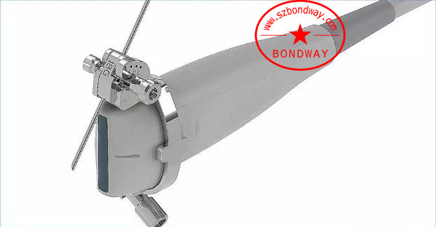
Pic 4.1 Biopsy needle guide for 4D volume probes (For GE RAB6-D)
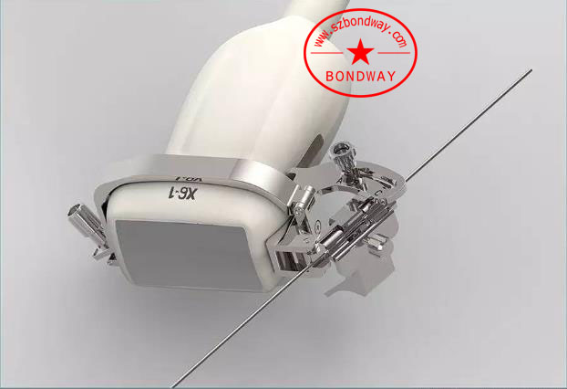
Pic 4.2 Biopsy needle guide for phased array transducer (For PHILIPSX6-1)
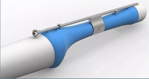
Pic 4.3 Biopsy needle guide for 4D volume endocavity transducer (For GE RIC5-9-D)
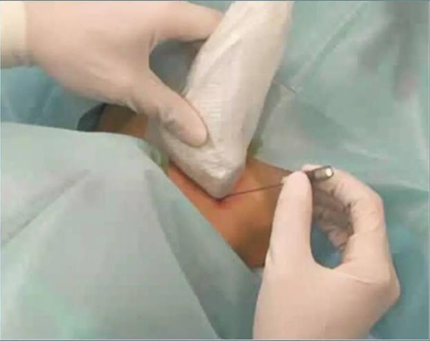
Pic 5 Freehand puncture of thyroid cells (using eight-beam suction biopsy needle)
3.1 PICC intervention application
Central venous catheter placement is an important medical technology widely used at present. It has been clinically used for more than 50 years and has formed a set of standardized procedures. In the fields of surgical anesthesia, intensive care treatment, emergency medicine, chemotherapy, hemodialysis, cardiovascular interventional therapy, bone marrow transplantation, high-energy nutritional support, and cardiopulmonary cerebral resuscitation, the application of central venous catheterization or central venous access has become basic in medical practice【3】
Central venipuncture catheterization requires a central venous puncture approach, which is based on the purpose of central venous catheterization, the patient’s own conditions (such as disease, physique, and co-existing diseases), and the puncture physician’s physiological knowledge and anatomy related to central venipuncture catheterization. Different veins and approaches are selected according to physical and anatomical knowledge, operation skills and clinical experience. Generally, there are subclavian veins, internal jugular veins, and expensive veins. It’s reported that traditional puncture techniques (without ultrasound guidance) can accidentally injure the arteries as high as 8% to 23% [4], and the application of modern ultrasound technology in central venous catheterization has greatly reduced the accidental injury rate of arteries.
The ultrasound-guided central venous puncture frame has two methods: in-plane and out-of-plane. As shown in Pic 6, both methods have their own advantages and disadvantages: the advantage of in-plane puncture is that the blind area is small and the needle insertion process can be seen in real time. The disadvantages are The needle insertion path is relatively long, which is not convenient for needle insertion in narrow areas; the advantage of out-of-plane is that the needle insertion path is short, and it is convenient for needle insertion even in narrow areas. The disadvantage is that the needle insertion is not visible, only when the puncture needle intersects the acoustic plane A bright spot will be displayed to show whether the puncture is in place.
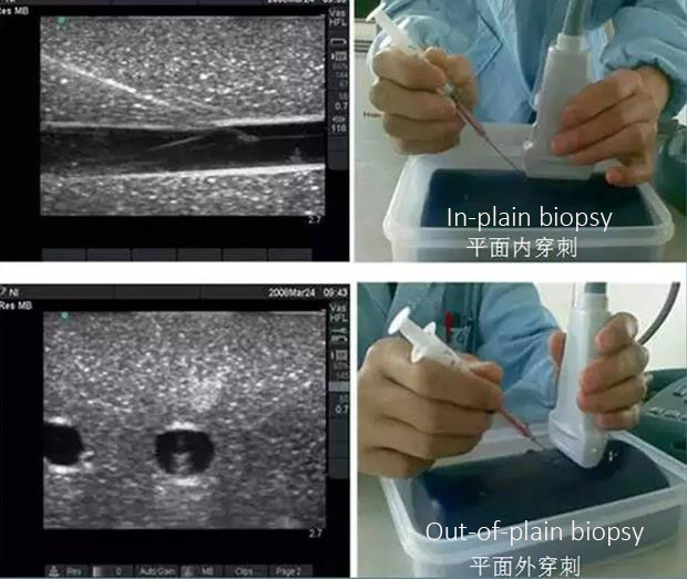
Pic 6 In-plain biopsy Vs Out-of-plain biopsy
As of the operators of central venous puncture, most of them are nursing staff. Regardless of in-plane or out-of-plane puncture, most cases are guided by biopsy needle guide. Pic 7 shows the same probe model with different puncture guidance methods. As shown in Pic 8, in order to meet the PICC out-of-plane puncture requirements of various ultrasound companies, a plastic fixing frame is set between the probe and the puncture needle guide block.
Pic 7.1 In-plain Vs Out-of-plain stainless steel biospy needle bracket
Pic 7.2 In-plain Vs Out-of-plain stainless steel biospy needle bracket
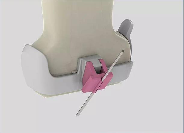
Pic 8.1 Plastic out-of-plain biospsy needle bracket (For Mindray 75L38EA)
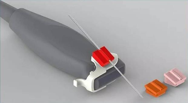
Pic 8.2 Plastic out-of-plain biospsy needle bracket (For Sonosite L25)
3.2 Application of abdominal intervention
The application of abdominal intervention mainly includes diagnostic and therapeutic puncture of liver, gallbladder, pancreas, spleen and kidney, catheter drainage, thermal ablation, etc. The following uses liver as an example to illustrate the application of biopsy needle bracket in abdominal intervention.
The interventional application of liver is mainly for the biopsy and treatment of solid liver tumors and the diagnosis and treatment of liver cysts. The puncture of the liver is generally in a relatively deep part. Therefore, most punctures are performed under the guidance of a large-sized convex probe or a micro-convex probe (Pic 9). Some clinical experts prefer to use phased array transducer. The probe is punctured (Pic 10).
Pic 9.1 Abdominal convex arry probe (For GE C1-5-D)
Pic 9.2 Abdominal micro-convex probe (For Hitachi EUP-B512)

Pic 10.1 Phased array probe with biospy needle guide (For Siemens 4V1)

Pic 10.2 Phased array probe with biospy needle guide (For GE S1-5)
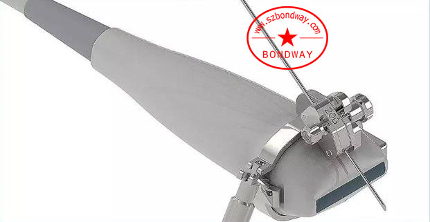
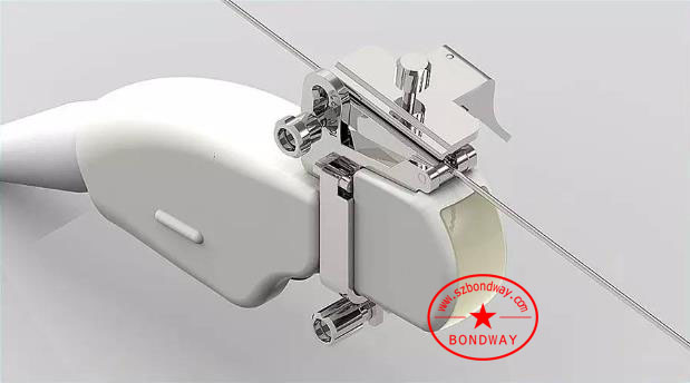
Because the interventional application of the liver includes histological biopsy (using thick needles ), cytological biopsy (using fine needles), aspiration and sclerotherapy of cysts (using fine needles), and thermal ablation treatment of solid tumors (using thick needles) , Cyst catheterization and drainage (use 6F-14F), the diameter of the needle or drainage tube used may be different specifications from 14-23G, 6F-14F, etc. Therefore, the range of the needle size of a needle guide is preferably adjustable. As shown in Pic 11, the size of the puncture needle track can be adjusted by rotating the adjusting nut to adapt to different needle types:
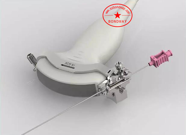
Pic 11 Biopsy needle guide with 14-22G needle (For Mindray SC5-1U)
3.3 Urinary intervention application
Prostate cancer is a malignant tumor that usually occurs in the epithelial cells in the prostate gland. At present, ultrasound-guided needle biopsy mainly has two routes: transrectal biopsy (TRBx) and transperineal Biopsy (TPBx). Through a comparative study of 339 patients, the ultrasound team of the Shanghai No. 10 Hospital concluded that the two puncture methods of TPBx and TRBx have the same effect in the diagnosis of prostate cancer, but the different needle insertion modes determine that the two punctures have different advantages. The disadvantages are as follows [5]:
1. The operation of TPBx is more complicated and requires higher requirements for the surgeon. The risk of sample failure is greater, and the pain experience of the patient during the operation is more significant (Pic 12)
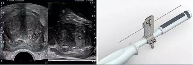
Pic 12 transperineal biospy needle bracket (For Mindray 6LB7)
2. The TRBx operation time is short and the pain during the operation is well tolerated. However, attention should be paid to avoid damaging the rectal wall and bladder during the operation, and to reduce serious postoperative complications such as rectal bleeding and infection. (Pic 13)
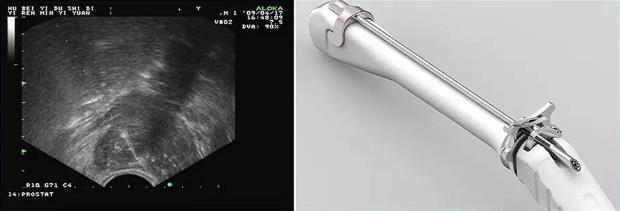
Pic 13 Transrectal biopsy needle bracket (For ALOKA UST-984-5)
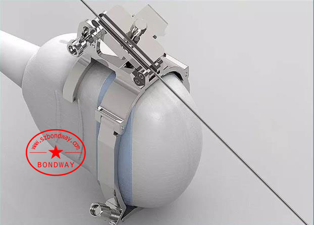
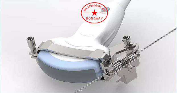
In the urinary interventional application, the puncture of the kidney is a very important direction. The common clinical applications include: extracorporeal shock wave lithotripsy under dual positioning of ultrasound and X-ray, ultrasound-guided minimally invasive percutaneous nephrolithotomy, ultrasound Guided minimally invasive percutaneous nephrostomy, ultrasound interventional treatment of renal cysts and polycystic kidneys. In the above puncture process, the dedicated puncture probe is preferred by clinicians because of its no-blind-zone and short needle path. Pic 14 shows the Esaote’s golf-shaped probe and biopsy needle guide.
Pic 14 Needle guide with needle inserted centrally (For ESAOTE SI2C41)
3.4 Interventional application of obstetrics and gynecology
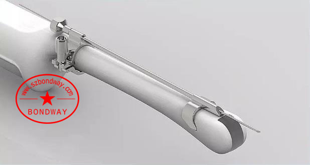
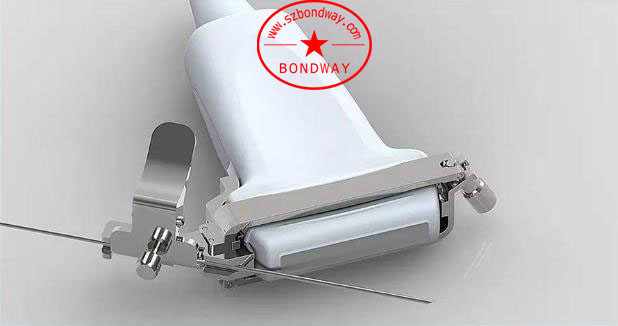
Interventional applications in obstetrics and gynecology are very extensive, including amniocentesis, cord blood puncture, ovarian cyst puncture, pelvic cyst puncture, and uterine fibroids puncture. Amniotic fluid puncture and cord blood puncture are generally performed under the guidance of a conventional abdominal probe and biopsy needle guide (see Pic 15); pelvic cysts and uterine fibroids are generally operated under the guidance of a transvaginal probe and biopsy needle guide (such as Pic 16) Sometimes, depending on the target position, puncture through the abdomen path is also used.
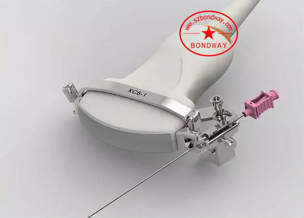
Pic 15 Biopsy needle guide attached onto Abdomenal convex probe (For SuperSonic XC6-1)

Pic 16 Biopsy needle guide mounted onto transvaginal probe (For PHILIPS C10-4ec)
3.5 Reproductive intervention applications
The application of ultrasound in reproductive intervention is mainly to perform follicular puncture with the vaginal probe and biopsy needle bracket, as shown in Pic 17 and Pic 18.

Pic 17 Biopsy needle bracket mounted onto endocavity transducer for egg retrieval purpose (For ALOKA UST-984-5)
Pic 18 Biopsy needle bracket mounted onto endocavity transducer for egg retrieval purpose (For Toshiba PVT-781VT)
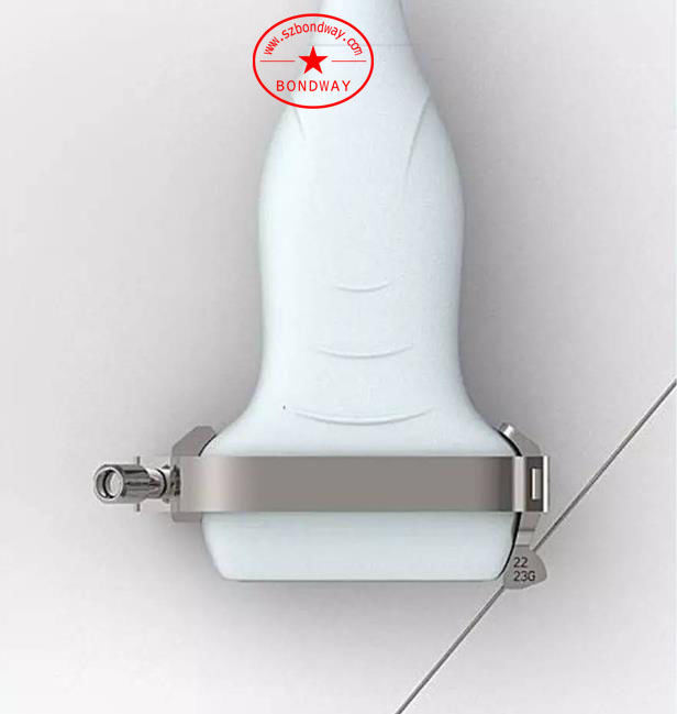
Pic 19 Biopsy needle guide with fixed needle track for high-frequency superficial transducer (For SonoSite HLF38x)
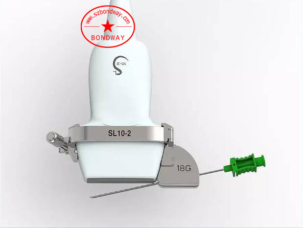
Pic 20 Biopsy needle guide with open-angle needle track for high-frequency superficial transducer (For SuperSonic SL10-2)
Pic 21 Biopsy needle guide with fine needle for thyroid biopsy purpose
Due to its non-radioactive, real-time, low price and other advantages, ultrasound has been accepted and recognized by more and more clinical departments and patients. Therefore, ultrasound interventional applications are not only widely used in the above-mentioned clinics, but also in pain management, nerve Block, orthopedics, cardiovascular disease, neurology and surgery, hematology, ENT, etc.
Shenzhen Bondway Electronics Co., Ltd. has developed over 600 types of reusable biospy needle brackets (or stainless steel biopsy needle guides) over the past few years, covering all world leading ultrasound brands, and will continue to offer innovative biopsy needle guidance products in the years to come. Please donot hesitate to contact us once you’ve any enquiries.

Best sellers
-
 Digital Vet Ultrasound scanner for equine, bovine, cattle, llama
-
 Veterinary ultrasound machine for equine, bovine,camel
-
 Digital Palmtop veterinary ultrasound scanner (Discontinued since April 29, 2022)
-
 Digital veterinary ultrasound for farm animals
-
 Digital Veterinary Ultrasound Scanner-ultrasound scan for swine, obvine,goat,alpacca
-
副本.png?imageView2/2/w/200/h/150/q/60) Digital color doppler ultrasound imaging system
Digital color doppler ultrasound imaging system
-
 Digital color doppler ultrasound imaging system
-
 Digital color doppler ultrasound imaging system


副本.png?imageView2/2/w/200/h/150/q/60)