 English
 中文
中文 Français
 Español
 Pусский
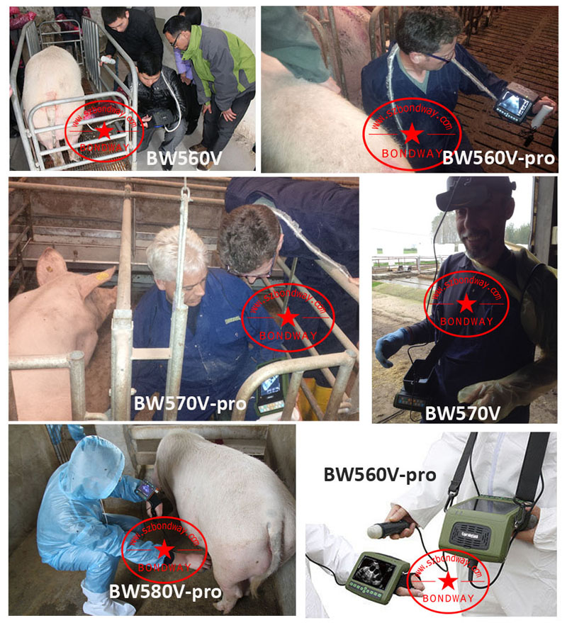
Using real-time veterinary ultrasound for pregnancy in swine, ultrasound scan for pig
What an ultrasound scan for pig can help
Sows that fail to establish and maintain pregnancy fail to cover costs associated with their daily maintenance and housing. Swine Ultrasound Pregnancy diagnosis can help to:
1) minimize costs associated with nonproductive days (NPDs),
2) maintain correct number of sows for farrowing crates,
3) identify open females for rebreeding or culling,
4) prevent unintended culling of pregnant sows,
5) identify the timing and extent of reproductive failure
6) help predict future pig flow
The Records concerned indicate herds average 70 NPDs with 20% due mated sows that fail to farrow. Of these failures, 40% return to estrus at about 21 days, while veterinary ultrasound can be used to identify an additional 35% between days 24 to 35. The remaining 25% of failures, lose their pregnancy after these days. Early identification of non-pregnant animals can facilitate rapid rebreeding or timely removal to maximize their value as cull sows.
Principles of ultrasound imaging system
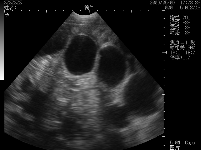
Real-time B-mode (brightness mode) ultrasonography displays a 2-dimensional image in gray scale. The image is composed of dots, that vary from white to light gray for very dense tissues such as the uterus and skin, and from dark gray to black for fluids and less dense tissues. For pregnancy diagnosis, decisions are based on the appearance of fluid vesicles (black) within the surrounding uterine tissue
Real-time ultrasound is based on the ability of specialized crystals within a transducer to vibrate and emit ultrasonic waves when an electric current is applied. Certain characteristics of ultrasound influence its ability to produce an accurate image for pregnancy diagnosis. For example, an ultrasonic wave is characterized by the distance it travels (wavelength), and the number of times the wave repeats within a second (frequency). The size of the crystal determines the wave, and the larger the crystals, the longer the wavelength and the lower the frequency. Larger crystals such as the 3.5 MHz, produce low frequency ultrasound waves that penetrate deep into the soft tissues of the animal. However, they provide lower image resolution (ability to distinguish between different structures) since fewer waves return and since more are lost waves over the increased distance traveled. In contrast, the smaller crystals of the 5.0-7.5 MHz transducers produce signals that travel shorter distances, but produce higher image resolution, since fewer waves are lost. These characteristics allow choices to be made for selection of transducers, since one will provide greater depth penetration but with lower resolution, while the other will facilitate shallow imaging, but with higher resolution. The correct choice of transducer involves the design of the equipment, the cost, and the anatomical depth of the structures to be visualized.
Figure 1. Representative trans-abdominal ultrasound images of a day 30 pregnant sow using a 3.5 MHz sector transducer. In each image, the multiple, irregular black shaped structures are the fluid-filled embryonic vesicles surrounded by the dense tissues of the uterus.
Equipment Selection
Most ultrasound systems is consisted of the main unit and the transducer. The transducer houses the crystals and is the only piece of equipment that is applied to the animal. Choosing one correct ultrasound scan for swine must take into account the practical aspects for its intended use.
For example
Many swine gestation stalls, with access only by long, narrow alleyways, and with limited electrical outlets. This makes imaging with large, heavy equipment difficult to accomplish. And non-portable equipment is impractical except when imaging can be performed in a centralized scanning area.
The portable veterinary ultrasound are designed for use in modern swine facilities and contain many of the features of the larger more expensive units. The portability classification arises from their design for mobility, which includes the unit weight, durability, and self-contained batteries. In general, they provide good quality imaging, and most have some additional features such as image enhancing or image storage capability. When examining these machines, practical ease of use in the barns must be evaluated. The ease of screen visualization should be considered since reflection of light off the screen is a common problem and makes fast diagnosis more challenging. In addition, access to the control panel, and ease of image adjustment should be tested, since image adjustment is often required between animals.
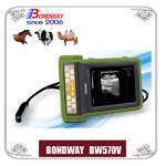
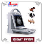
BW520V, BW540V, BW560V, BW570V, BW560V-pro, BW570V-pro and BW580V-pro etc are durable, reliable, made for the rugged farm environment and ideal for swine reproduction scanning.
Shenzhen Bondway Electronics Co.Ltd
2018-7-25
Best sellers
-
 Digital Vet Ultrasound scanner for equine, bovine, cattle, llama
-
 Veterinary ultrasound machine for equine, bovine,camel
-
 Digital Palmtop veterinary ultrasound scanner (Discontinued since April 29, 2022)
-
 Digital veterinary ultrasound for farm animals
-
 Digital Veterinary Ultrasound Scanner-ultrasound scan for swine, obvine,goat,alpacca
-
副本.png?imageView2/2/w/200/h/150/q/60) Digital color doppler ultrasound imaging system
Digital color doppler ultrasound imaging system
-
 Digital color doppler ultrasound imaging system
-
 Digital color doppler ultrasound imaging system


副本.png?imageView2/2/w/200/h/150/q/60)